Introduction and Objective: Unlike other LA users, most Elekta users use a 2D Array or other external devices for Patient specific QA purpose. In India, we experimented with a third-party portal dosimetry system called EPI Beam from DosiSoft, France, for PSQA. EPI (Electronic Portal Imaging) Beam Portal Dosimetry is a method used to verify the dose delivery in radiation therapy. It utilizes the Elekta iViewGT EPID to measure the radiation dose distribution delivered to the patient. The acquired portal images are then compared with the predicted dose distribution from the treatment planning system (TPS). The objective of this study is to analyze the gamma pass rate with this technology in various sites and treatment techniques in 100 patients. Material and methods: Pre-treatment verification using Portal Dosimetry was performed on 100 patients utilizing an Elekta iViewGT™ EPID and EPI Beam software on an Elekta Harmony Pro linear accelerator having 6 MV, 15 MV, and 6FFF photon energies. Elekta’s portal dosimetry system eliminates the need for a separate QA plan or a phantom, as the patient's treatment plan is directly delivered to the Electronic Portal Imaging Device (EPID) panel. The delivered dose is then calculated and compared to the planned dose, ensuring accurate dose delivery and proper linear accelerator functioning before the patient’s first treatment fraction. Results: In all plans that were done by Volumetric Modulated Arc Therapy (VMAT) for various diagnoses were analyzed and the average gamma pass rate for Head and Neck, Thoracic, Pelvis, Breast, are 99.34%, 98.97%, 98.78% and 99.35% respectively. Conclusions: The gamma evaluation indicates a good correlation between predicted and acquired EPID image doses. The EPID-based pre-treatment verification using EPI Beam from DosiSoft is a time-saving and comfortable tool for performing pre-treatment verification. This method enhances the precision and safety of cancer treatments.
| Published in | American Journal of Clinical and Experimental Medicine (Volume 13, Issue 1) |
| DOI | 10.11648/j.ajcem.20251301.11 |
| Page(s) | 1-7 |
| Creative Commons |
This is an Open Access article, distributed under the terms of the Creative Commons Attribution 4.0 International License (http://creativecommons.org/licenses/by/4.0/), which permits unrestricted use, distribution and reproduction in any medium or format, provided the original work is properly cited. |
| Copyright |
Copyright © The Author(s), 2025. Published by Science Publishing Group |
EPID, Portal Dosimetry, Epibeam, Elekta Medical Systems, Radiation Oncology, Patient Specific Quality Assurances, Pre-Treatment Verification
Gamma analysis results of commissioning verification of three QA plans for 6 MV | |||||
|---|---|---|---|---|---|
Field | DTA (mm) | DD (%) | GAI (%) | Energy | |
1 | E field, | 2 | 2 | 100 | 6MV |
2 | Triangle field | 2 | 2 | 100 | 6MV |
3 | chevron field | 2 | 2 | 100 | 6MV |
1 | E field, | 2 | 2 | 98.95 | 6MV FFF |
2 | Triangle field | 2 | 2 | 100 | 6MV FFF |
3 | chevron field | 2 | 2 | 100 | 6MV FFF |
1 | E field, | 2 | 2 | 100 | 15MV |
2 | Triangle field | 2 | 2 | 100 | 15MV |
3 | chevron field | 2 | 2 | 100 | 15MV |
EPID | Electronic Portal Imaging Device |
PSQA | Patient Specific Quality Assurance |
Epibeam | Electronic Portal Imaging Beam |
VMAT | Volumetric Modulated Arc Therapy |
SBRT | Steriotactic Body Radiotherapy |
| [1] | Mikel Byrne, Ben Archibald-Heeren, Yunfei Hu, Andrew Fong, Leena Chong, Amy Teh. Comparison of semiautomated tangential VMAT with 3DCRT for breast or chest wall and regional nodes. Journal of applied clinical Medical Physic. 2018, 19: 5: 684-693. |
| [2] | Clements, M.; Schupp, N.; Tattersall, M.; Brown, A.; Larson, R. Monaco treatment planning system tools and optimization processes. Med. Dosim. 2018, 43, 106-117. |
| [3] | Malouff, T. D.; Seneviratne, D.; Stross, W. C.; Ko, S.; Tzou, K.; Trifiletti, D. M.; Vallow, L. A. Public interest in stereotactic body radiation therapy (SBRT) and stereotactic radiosurgery (SRS) in the United States. J. Radiosurg. SBRT. 2020, 6, 311-315. |
| [4] | Saito, M.; Komiyama, T.; Marino, K.; Aoki, S.; Oguri, M.; Yamada, T.; Sano, N.; Suzuki, H.; Ueda, K.; Onishi, H. Dosimetric effects of differences in multi-leaf collimator speed on SBRT-VMAT for central lung cancer patients. Technol. Cancer Res. Treat. 2022, 21, 15330338221119752. |
| [5] | Kruszyna, M.; Skrobala, A.; Romanski, P.; Ryczkowski, A.; Suchorska, W.; Kulcenty, K.; Piotrowski, I.; Borowicz, D.; Graczyk, K.; Matuszak, N.; et al. Influence of Specific Treatment Parameters on Nontarget and Out-of-Field Doses in a Phantom Model of Prostate SBRT with CyberKnife and TrueBeam. Life 2022, 12, 628. |
| [6] | Serra, M.; de Martino, F.; Savino, F.; D’Alesio, V.; Arrichiello, C.; Quarto, M.; Loffredo, F.; di Franco, R.; Borzillo, V.; Muto, M.; et al. SBRT for Localized Prostate Cancer: CyberKnife vs. VMAT-FFF, a Dosimetric study. Life 2022, 12, 711. |
| [7] | Ezzell GA, Galvin JM, Low D, et al. Guidance document on delivery, treatment planning, and clinical implementation of IMRT: report of the IMRT subcommittee of the AAPM Radiation Therapy Committee. Med Phys. 2003; 30(8): 2089-115. |
| [8] | Hartford AC, Galvin JM, Beyer DC, et al. American College of Radiology (ACR) and American Society for Radiation Oncology (ASTRO) practice guideline for intensity-modulated radiation therapy (IMRT). Am J Clin Oncol. 2012; 35(6): 612-17. |
| [9] | Van Esch A, Huyskens DP, Hirschi L, Baltes C. Optimized Varian aSi portal dosimetry: development of datasets for collective use. J Appl Clin Med Phys. 2013; 14(6): 82-99. |
| [10] | Vinall AJ, Williams AJ, Currie VE, Van Esch A, Huyskens D. Practical guidelines for routine intensity-modulated radiotherapy verification: pre-treatment verification with portal dosimetry and treatment verification with in vivo dosimetry. Br J Radiol. 2010; 83(995): 949-57. |
| [11] | Li, J. G.; Yan, G.; Liu, C. Comparison of two commercial detector arrays for IMRT quality assurance. J. Appl. Clin. Med. Phys. 2009, 10, 62-74. |
| [12] | Gloi, A. M.; Buchana, R. E.; Zuge, C. L.; Goettler, A. M. RapidArc quality assurance through MapCHECK. J. Appl. Clin. Med. Phys. 2011, 12, 3251. |
| [13] | Syamkumar, S. A.; Padmanabhan, S.; Sukumar, P.; Nagarajan, V. Characterization of responses of 2d array seven29 detector and its combined use with octavius phantom for the patient-specific quality assurance in rapidarc treatment delivery. Med. Dosim. 2012, 37, 53-60. |
| [14] | Diaz Moreno, R. M.; Venencia, D.; Garrigo, E.; Pipman, Y. A method to enhance 2D ion chamber array patient specific quality assurance for IMRT. Australas. Phys. Eng. Sci. Med. 2017, 40, 145-151. |
| [15] | Herwiningsih, S.; Hanlon, P.; Fielding, A. Sensitivity of an Elekta iView GT a-Si EPID model to delivery errors for pre-treatment verification of IMRT fields. Australas. Phys. Eng. Sci. Med. 2014, 37, 763-770. |
| [16] | Seung-Hyeop Baek, Sang-Hyoun Choi, Moo-Jae Han, Gyu-Seok Cho, Wonil Jang, Jin-Sung Kim and Kum-Bae Kim. Clinical Efficacy of an Electronic Portal Imaging Device versus a Physical Phantom Tool for Patient-Specific Quality Assurance, Life 2022, 12, 1923. |
| [17] | Shalev S. On-line portal imaging: Contributions and limitations in clinical practice. Front Radiat Ther Oncol. 1996; 29: 156-167. |
| [18] | Herman MG, Kruse JJ, Hagness CR. Guide to clinical use of electronic portal imaging. J Appl Clin Med Phys. 2000; 1: 38-57. |
| [19] | Gądek A, Plaza D, Sroka Ł, Reudelsdorf-Ullmann M, Ślosarek K. Comparison of EPID portal dosimetry verification and RadCalc dose verification for VMAT treatment plans. NOWOTWORY J Oncol 2024; 74: 12-19. |
| [20] | Ivan Kutuzov, Ryan Rivest, Eric VanUytven, Boyd McCurdy. Long-term performance monitoring of a-Si 1200 electronic portal imaging device for dosimetric applications. |
APA Style
Muralidhar, K. R., Nishaanth, B., Kumar, A. V., Srinivas, P., Kolluru, V. R. R., et al. (2025). Clinical Application of Portal Dosimetry for Pre-treatment Dosimetric Verification Across Various Diagnoses Using Electronic Portal Imaging Integrated with Epibeam. American Journal of Clinical and Experimental Medicine, 13(1), 1-7. https://doi.org/10.11648/j.ajcem.20251301.11
ACS Style
Muralidhar, K. R.; Nishaanth, B.; Kumar, A. V.; Srinivas, P.; Kolluru, V. R. R., et al. Clinical Application of Portal Dosimetry for Pre-treatment Dosimetric Verification Across Various Diagnoses Using Electronic Portal Imaging Integrated with Epibeam. Am. J. Clin. Exp. Med. 2025, 13(1), 1-7. doi: 10.11648/j.ajcem.20251301.11
AMA Style
Muralidhar KR, Nishaanth B, Kumar AV, Srinivas P, Kolluru VRR, et al. Clinical Application of Portal Dosimetry for Pre-treatment Dosimetric Verification Across Various Diagnoses Using Electronic Portal Imaging Integrated with Epibeam. Am J Clin Exp Med. 2025;13(1):1-7. doi: 10.11648/j.ajcem.20251301.11
@article{10.11648/j.ajcem.20251301.11,
  author = {Kanaparthy Raja Muralidhar and Balasubramaniyam Nishaanth and Akkupalli Vijay Kumar and Ponaganti Srinivas and Veera Raghavendra Rao Kolluru and Rampally Kumar and Venkataramanan Ramachandran},
  title = {Clinical Application of Portal Dosimetry for Pre-treatment Dosimetric Verification Across Various Diagnoses Using Electronic Portal Imaging Integrated with Epibeam
},
  journal = {American Journal of Clinical and Experimental Medicine},
  volume = {13},
  number = {1},
  pages = {1-7},
  doi = {10.11648/j.ajcem.20251301.11},
  url = {https://doi.org/10.11648/j.ajcem.20251301.11},
  eprint = {https://article.sciencepublishinggroup.com/pdf/10.11648.j.ajcem.20251301.11},
  abstract = {Introduction and Objective: Unlike other LA users, most Elekta users use a 2D Array or other external devices for Patient specific QA purpose. In India, we experimented with a third-party portal dosimetry system called EPI Beam from DosiSoft, France, for PSQA. EPI (Electronic Portal Imaging) Beam Portal Dosimetry is a method used to verify the dose delivery in radiation therapy. It utilizes the Elekta iViewGT EPID to measure the radiation dose distribution delivered to the patient. The acquired portal images are then compared with the predicted dose distribution from the treatment planning system (TPS). The objective of this study is to analyze the gamma pass rate with this technology in various sites and treatment techniques in 100 patients. Material and methods: Pre-treatment verification using Portal Dosimetry was performed on 100 patients utilizing an Elekta iViewGT™ EPID and EPI Beam software on an Elekta Harmony Pro linear accelerator having 6 MV, 15 MV, and 6FFF photon energies. Elekta’s portal dosimetry system eliminates the need for a separate QA plan or a phantom, as the patient's treatment plan is directly delivered to the Electronic Portal Imaging Device (EPID) panel. The delivered dose is then calculated and compared to the planned dose, ensuring accurate dose delivery and proper linear accelerator functioning before the patient’s first treatment fraction. Results: In all plans that were done by Volumetric Modulated Arc Therapy (VMAT) for various diagnoses were analyzed and the average gamma pass rate for Head and Neck, Thoracic, Pelvis, Breast, are 99.34%, 98.97%, 98.78% and 99.35% respectively. Conclusions: The gamma evaluation indicates a good correlation between predicted and acquired EPID image doses. The EPID-based pre-treatment verification using EPI Beam from DosiSoft is a time-saving and comfortable tool for performing pre-treatment verification. This method enhances the precision and safety of cancer treatments.
},
 year = {2025}
}
TY  - JOUR
T1  - Clinical Application of Portal Dosimetry for Pre-treatment Dosimetric Verification Across Various Diagnoses Using Electronic Portal Imaging Integrated with Epibeam

AU  - Kanaparthy Raja Muralidhar
AU  - Balasubramaniyam Nishaanth
AU  - Akkupalli Vijay Kumar
AU  - Ponaganti Srinivas
AU  - Veera Raghavendra Rao Kolluru
AU  - Rampally Kumar
AU  - Venkataramanan Ramachandran
Y1  - 2025/02/07
PY  - 2025
N1  - https://doi.org/10.11648/j.ajcem.20251301.11
DO  - 10.11648/j.ajcem.20251301.11
T2  - American Journal of Clinical and Experimental Medicine
JF  - American Journal of Clinical and Experimental Medicine
JO  - American Journal of Clinical and Experimental Medicine
SP  - 1
EP  - 7
PB  - Science Publishing Group
SN  - 2330-8133
UR  - https://doi.org/10.11648/j.ajcem.20251301.11
AB  - Introduction and Objective: Unlike other LA users, most Elekta users use a 2D Array or other external devices for Patient specific QA purpose. In India, we experimented with a third-party portal dosimetry system called EPI Beam from DosiSoft, France, for PSQA. EPI (Electronic Portal Imaging) Beam Portal Dosimetry is a method used to verify the dose delivery in radiation therapy. It utilizes the Elekta iViewGT EPID to measure the radiation dose distribution delivered to the patient. The acquired portal images are then compared with the predicted dose distribution from the treatment planning system (TPS). The objective of this study is to analyze the gamma pass rate with this technology in various sites and treatment techniques in 100 patients. Material and methods: Pre-treatment verification using Portal Dosimetry was performed on 100 patients utilizing an Elekta iViewGT™ EPID and EPI Beam software on an Elekta Harmony Pro linear accelerator having 6 MV, 15 MV, and 6FFF photon energies. Elekta’s portal dosimetry system eliminates the need for a separate QA plan or a phantom, as the patient's treatment plan is directly delivered to the Electronic Portal Imaging Device (EPID) panel. The delivered dose is then calculated and compared to the planned dose, ensuring accurate dose delivery and proper linear accelerator functioning before the patient’s first treatment fraction. Results: In all plans that were done by Volumetric Modulated Arc Therapy (VMAT) for various diagnoses were analyzed and the average gamma pass rate for Head and Neck, Thoracic, Pelvis, Breast, are 99.34%, 98.97%, 98.78% and 99.35% respectively. Conclusions: The gamma evaluation indicates a good correlation between predicted and acquired EPID image doses. The EPID-based pre-treatment verification using EPI Beam from DosiSoft is a time-saving and comfortable tool for performing pre-treatment verification. This method enhances the precision and safety of cancer treatments.

VL  - 13
IS  - 1
ER  -
Karkinos Healthcare, Hyderbad, India
Karkinos Healthcare-CIMS, Bhopal, India
Karkinos Healthcare-Udayananda, Nandyala, India
Karkinos Healthcare, Hyderbad, India
Karkinos Healthcare, Hyderbad, India
Karkinos Healthcare, Hyderbad, India
Karkinos Healthcare, Mumbai, India
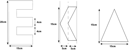
Figure 1. Pre-Treatment beam data templates.
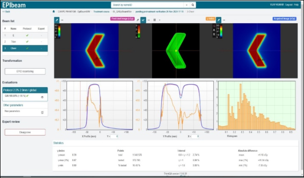
Figure 2. Gamma Analysis of Chevron Field in Epibeam.
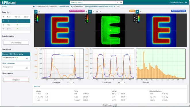
Figure 3. Gamma Analysis of E Field in Epibeam.
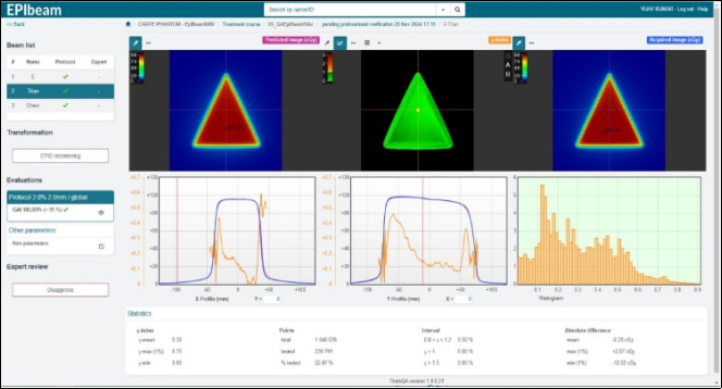
Figure 4. Gamma Analysis of Triangle Field in Epibeam.
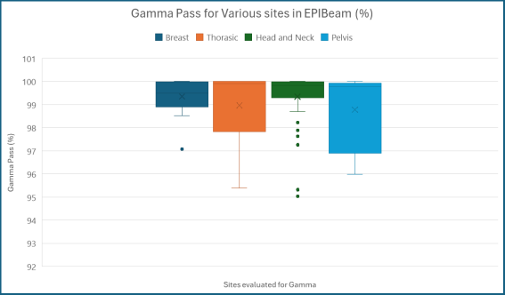
Figure 5. Gamma Pass Index for Breast, Thorax, Head and Neck and Pelvis Cases.
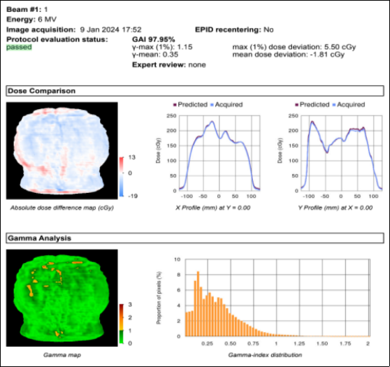
Figure 6. PSQA in EPIbeam in Elekta Harmony Pro Linear accelerator.